
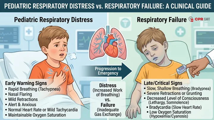
Recognizing respiratory compromise early is one of the most critical skills taught in Pediatric Advanced Life Support (PALS). Unlike adults, children frequently present with breathing problems long before they experience cardiovascular collapse. Identifying a worsening respiratory status creates a vital window of opportunity for intervention before a critical event occurs.
One of the most important clinical distinctions healthcare providers must master is the difference between respiratory distress and respiratory failure. Although the terms are sometimes used interchangeably in casual conversation, they represent entirely different physiological states. The ability to recognize exactly where a child falls on this clinical continuum dictates treatment decisions, urgency, and patient outcomes.
Quick Answer: What Is the Difference between Pediatric Respiratory Distress and Respiratory Failure?
Pediatric respiratory distress occurs when a child experiences an increased work of breathing but can still maintain adequate oxygenation and ventilation. The body is successfully compensating by recruiting additional mechanisms (like faster breathing or accessory muscle use) to preserve respiratory function.
Pediatric respiratory failure occurs when the child can no longer maintain adequate oxygenation or ventilation despite compensatory effort. The respiratory system can no longer sustain adequate oxygenation, ventilation, or both. Immediate, aggressive intervention becomes necessary.
The key difference is simple: a child in respiratory distress is compensating, while a child in respiratory failure is actively decompensating.
Why Pediatric Patients Deteriorate Differently
Children do not experience respiratory compromise in the same way adults do. Their distinct anatomy and physiology create unique challenges that directly influence how symptoms develop and progress.
Children have smaller airways, meaning even a millimeter of swelling or minor mucus obstruction creates substantial resistance to airflow. Furthermore, pediatric patients possess significantly higher metabolic demands and consume oxygen much more rapidly than adults. Because their chest walls are highly compliant (pliable) and their respiratory reserve is limited, infants and young children are uniquely vulnerable to rapid deterioration.
Because of these differences, pediatric patients often compensate incredibly well during the early stages of an illness. A child may appear relatively stable despite exerting massive respiratory effort. However, once muscle fatigue develops and their compensatory mechanisms fail, deterioration can occur rapidly once compensation fails. This physiological pattern explains the classic pediatric emergency scenario: a child who appears stable for hours, only to suddenly “crash” and become critically ill.
Understanding Pediatric Respiratory Distress
Respiratory distress develops when a child encounters a barrier to normal breathing but manages to maintain oxygenation and ventilation through increased physical effort.
Many common pediatric illnesses trigger respiratory distress. Conditions such as asthma exacerbations, bronchiolitis, pneumonia, severe allergic reactions, and upper airway obstructions force children to work much harder to breathe. Fever and systemic metabolic stress can also increase respiratory demand.
During this stage, increased effort may temporarily preserve blood oxygen and carbon dioxide levels, but this compensation is physically exhausting and cannot continue indefinitely. Without treatment, respiratory muscle fatigue is inevitable.
Clinical Signs of Respiratory Distress
The most obvious feature of respiratory distress is the visible increased work of breathing. Healthcare providers must assess how hard the child is working rather than relying on a single vital sign.
- Tachypnea: Breathing faster is often the earliest sign, as the child attempts to maintain oxygen delivery.
- Nasal Flaring: Infants and younger children frequently flare their nostrils to intuitively decrease airway resistance and maximize airflow.
- Retractions: As respiratory effort increases, the use of accessory muscles becomes visible. Intercostal (between ribs), substernal (below the breastbone), supraclavicular (above the collarbones), and subcostal retractions all indicate severe effort. Infants may also demonstrate “head bobbing” caused by the vigorous use of neck muscles during inhalation.
- Audible Airway Sounds: Wheezing generally suggests lower airway disease, stridor indicates upper airway obstruction, and crackles point toward pulmonary fluid or pneumonia.
Crucially, children in respiratory distress frequently remain alert. Anxiety, restlessness, and irritability are incredibly common because the child is hyper-aware of their breathing difficulty, yet they are still maintaining adequate oxygen delivery to the brain.
Understanding Pediatric Respiratory Failure
Respiratory failure marks the dangerous turning point where compensatory mechanisms collapse. The respiratory system can no longer sustain the minimum required oxygenation or clear carbon dioxide effectively.
This stage may develop gradually as untreated respiratory distress progresses to exhaustion, or it can occur suddenly in severe disease processes like advanced asthma, neurologic impairment, sepsis, or toxic ingestion.
Clinical Signs of Respiratory Failure (The “Quiet Child”)
Respiratory failure often looks drastically different from respiratory distress. Instead of demonstrating extreme effort, children may actually appear calmer or less active. This presentation is highly deceptive.
As severe fatigue sets in, respiratory effort decreases. Breathing may become shallow, irregular, weak, or unusually slow (bradypnea). A child who previously exhibited dramatic retractions may suddenly show less visible work of breathing simply because their muscles are too exhausted to continue the fight.
Mental status changes are the most critical indicator during this stage. Lethargy, confusion, poor responsiveness to pain or parents, and decreased interaction indicate worsening cerebral hypoxia (lack of oxygen to the brain) and impaired systemic perfusion.
Furthermore, breath sounds often become diminished. In a severe asthma exacerbation, a “quiet chest” with minimal wheezing is not a sign of improvement; it is a red flag indicating that air movement has practically stopped. Central cyanosis (blue discoloration of the lips and core), prolonged apnea, and bradycardia are late, pre-arrest findings.
Distress vs. Failure: A Side-by-Side Comparison
| Clinical Finding | Respiratory Distress | Respiratory Failure |
| Respiratory Effort | Increased (retractions, flaring) | Decreased, shallow, or ineffective |
| Respiratory Rate | Usually fast (tachypnea) | Slow, weak, or irregular |
| Mental Status | Alert, anxious, or irritable | Altered, lethargic, or unresponsive |
| Skin Color | Usually normal | Pale, mottled, or cyanotic |
| Breath Sounds | Present (wheezes, stridor) | Diminished or entirely absent |
| Physiologic State | Compensation maintained | Compensation failing |
| Urgency | Serious; requires prompt care | Immediate life-saving intervention |
How PALS Approaches Pediatric Respiratory Emergencies
PALS methodology teaches a structured, systematic approach to identify respiratory compromise before it progresses to cardiac arrest.
Providers begin with the Initial Impression (often called the Pediatric Assessment Triangle), which evaluates appearance, work of breathing, and circulation to the skin. This rapid visual assessment provides an immediate snapshot of clinical severity and frequently reveals the earliest signs of instability.
Treatment strategies depend entirely on where the child falls on the continuum. During respiratory distress, interventions focus on supplemental oxygen, bronchodilator therapy, targeted medication, and close observation. Conversely, respiratory failure requires immediate escalation, often necessitating bag-mask ventilation or advanced airway placement.
When respiratory compromise progresses rapidly because of severe upper airway pathology, review our specialized clinical guide on Pediatric Airway Emergencies: Croup & Inhalation Injury.
Common Recognition Errors in Emergency Care
Delayed recognition typically occurs when providers focus on a single symptom rather than interpreting the entire clinical picture. Avoid these common diagnostic traps:
- Misinterpreting Fatigue as Improvement: Assuming decreased respiratory effort means the child is getting better is a deadly mistake. Decreasing effort combined with lethargy reflects muscle exhaustion and impending failure.
- Over-relying on Pulse Oximetry: Oxygen saturation does not define respiratory status. A child can have a falsely reassuring SpO2 of 95% while suffering from severe hypercarbia (retained carbon dioxide) and ventilation failure.
- Waiting for Cyanosis: Central cyanosis and bradycardia are late, ominous signs in pediatric patients. If you wait for a child to turn blue or their heart rate to drop before intervening, you are already dangerously close to cardiac arrest.
Frequently Asked Questions
What is the difference between respiratory distress and respiratory failure in children?
Respiratory distress occurs when a child works harder to maintain breathing but continues compensating effectively. Respiratory failure develops when the child becomes exhausted and the respiratory system can no longer adequately maintain oxygenation or ventilation.
Can respiratory distress progress into respiratory failure?
Yes. If the underlying cause is not treated, respiratory distress will eventually lead to muscle fatigue, worsening oxygenation, impaired ventilation, and respiratory failure.
Is pediatric respiratory failure considered an immediate emergency?
Absolutely. Respiratory failure requires immediate, aggressive intervention (such as positive pressure ventilation) because it is the one of the leading contributors to pediatric cardiac arrest.
Why is bradycardia highly concerning in pediatric respiratory emergencies?
Unlike adults who frequently experience primary cardiac events, children usually suffer from secondary cardiac arrest driven by lack of oxygen. Bradycardia (an abnormally slow heart rate) develops due to severe, prolonged hypoxia and is a late, pre-arrest sign of respiratory failure.
Why can a “quieter” child sometimes be more clinically concerning?
If a child who was previously agitated and struggling to breathe suddenly becomes quiet, lethargic, and exerts less effort, it usually indicates profound exhaustion and worsening cerebral hypoxia rather than clinical improvement.